Novel endomyocardial biopsy technique of porcine xenohearts in brain-dead decedents
Bernard Kadosh1, Daniel Bamira1, Tajinderpal Saraon1, Randal I Goldberg1, Michael DiVita1, Alex Reyentovich1, Jeffrey Stern1, Karen Khalil1, Jacqueline Kim1, Thomas Jan1, Jennie Ngai1, David Ayares2, Elaina Weldon1, Adam Griesemer1, Deane Smith1, Robert A Montgomery1, Nader Moazami1.
1NYU Langone Transplant Institute, New York, NY, United States; 2Revivicor, Inc., Blacksburg, VA, United States
Introduction: The objective of this experiment was to describe the endomyocardial biopsy technique in two brain-dead decedent porcine xenoheart transplant recipients.
Methods: Two 10-gene-edited porcine xenograft hearts were transplanted into brain-dead decedents and monitored over 66 hours. Endomyocardial biopsies were performed on POD 1 and POD 2 in order to surveil for hyperacute and acute xenograft rejection. In order to ensure proper positioning of the bioptome without damaging adjacent structures or perforating the heart, biopsies were performed under continuous transesophageal echocardiography guidance.
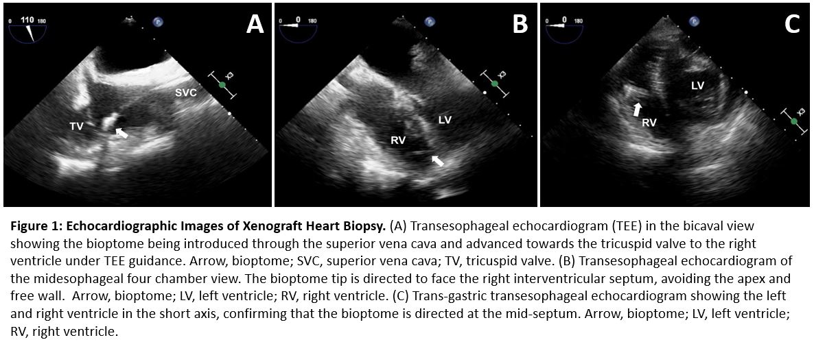
Results: An 8.5-Fr introducer sheath was used to access the right internal jugular vein. A 7-Fr, 50cm Argon Jawz Pre-Curved flexible bioptome was introduced several times through the 8.5-Fr sheath and advanced under echocardiographic guidance to the right ventricle. A bicaval view was used to guide the bioptome from the superior vena cava through the tricuspid valve (Fig 1A). Then, a 4-chamber view was used to guide tip of the bioptome to face the right interventricular septum, avoiding the apex and the right ventricular free wall (Fig 1B). Finally, a transgastric short axis view was used to ensure that the bioptome advanced into the mid-septum, avoiding the anterior and posterior septal insertion points (Fig 1C). 7 tissues samples were taken from the right mid-interventricular septum for analysis. No complications occurred during the study period.
Conclusion: Endomyocardial biopsy of porcine xenografts poses a unique challenge due to the morphological differences between human and porcine cardiac anatomy. The left ventricle is more dominant in porcine hearts, which shifts the septum to the right and limits the size of the right ventricular chamber. The moderator band is more prominent, limiting the ability to maneuver the bioptome freely. In addition, the external geometry of porcine hearts may alter their orientation in the human thorax after xenotransplantation. Conventional fluoroscopic guidance may be inadequate to ensure the safety of the procedure. The use of echocardiography is prudent to identify internal cardiac structures during endomyocardial biopsy of xenotransplanted hearts.
Supported by Lung Biotechnology, a wholly owned subsidiary of United Therapeutics..
References:
[1] Crick SJ, Sheppard MN, Ho SY, Gebstein L, Anderson RH. Anatomy of the pig heart: comparisons with normal human cardiac structure. J Anat. 1998 Jul; 193(Pt 1): 105–119. doi: 10.1046/j.1469-7580.1998.19310105.x
Lectures by Bernard Kadosh
| When | Session | Talk Title | Room |
|---|---|---|---|
|
Thu-26 14:45 - 15:45 |
Pre-clinical and clinical application of xenotransplantation and current barriers 1 | Novel Endomyocardial Biopsy Technique of Porcine Xenohearts in Brain-Dead Decedents | Indigo H |