Resident Physician
Cardiothoracic Surgery
University of Southern California
Preservation of cardiac xenografts in a model of infant human cardiac xenotransplantation
Chace Mitchell1, David C Cleveland1, David KC Cooper2, John Cleveland1.
1Division of Pediatric Cardiac Surgery, Heart Institute, Children’s Hospital of Los Angeles, Los Angeles, CA, United States; 2Center for Transplantation Sciences, Department of Surgery, Massachusetts General Hospital/Harvard Medical School, Boston, MA, United States
Introduction: There is no standard protocol for management of organ preservation for orthotopic, life-sustaining cardiac xenotransplantation, particularly for hearts from pediatric sized donors. Standard techniques and solutions successful in human allotransplantation are not viable. Published practices for cardiac xenotransplantation are cumbersome and expensive1,2. We theorized that a solution commonly used in reparative cardiac surgery in human children3 would suffice by exploiting the advantages inherent to xenotransplantation, namely the ability to reduce organ ischemic times by donor transport.
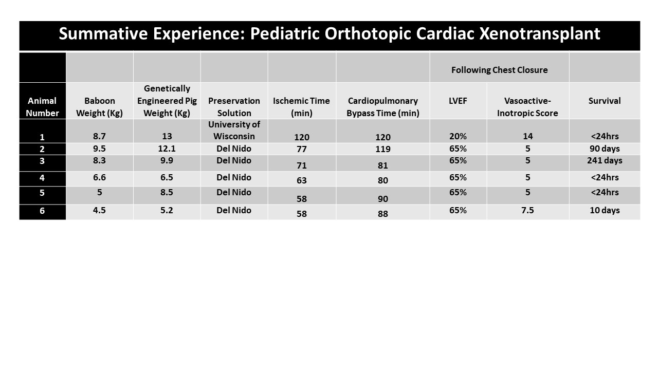
Method: We carried out 6 cardiac xenotransplants from genetically modified pigs to papio hamadryas baboons. Recipient weights ranged 4.4 to 9.5 kg, and age ranged from 13 to 24 months of age. The first porcine heart was prepared analogous to human allotransplant: University of Wisconsin (UW) solution for arrest and cold static preservation for 1 hour before transplant. The following 5 porcine hearts were preserved with a modified, hyper-oncotic Del Nido cardioplegia solution followed by immediate implant into our recipient. Post-operative echocardiograms, hemodynamics, and clinical outcomes were tracked.
Results: The first xenograft recipient preserved with UW weaned from bypass with marginal hemodynamics and poor cardiac function, expiring 3 hours post-operatively. Graft ischemic time was 120 minutes while cardiopulmonary bypass time was 120 minutes. The following 5 baboons preserved with Del Nido weaned from bypass on low dose inotropic support with normal cardiac function recorded on echocardiogram. Median graft ischemic time was 63 [58,71] minutes while median cardiopulmonary bypass time was 88 [81,90] minutes. Vasoactive-inotropic score was lower for the 5 baboons who received a heart preserved with Del Nido in comparison to the one preserved with UW. Of the 5 Del Nido baboons, three survived greater than 7 days post-operatively. Two went on to survive 90- and 241-days post-xenotransplantation.
Conclusion: As cardiac xenotransplant moves closer to clinical reality, it is necessary to develop standard practices that ensure the greatest chances of success. A failsafe method of cardiac xenograft preservation is an imperative component. Del Nido cardioplegia solution followed by immediate transplant demonstrates promising outcomes to avoid primary graft dysfunction for cardiac xenotransplantation in small animals.
References:
[1] Längin M, Mayr T, Reichart B, et al. Consistent success in life-support-ing porcine cardiac xenotransplantation. Nature. 2018;564:430–433.
[2] Matte GS, del Nido PJ. History and use of del Nido cardioplegia solution at Boston Children's Hospital. J Extra Corpor Technol. 2012 Sep;44(3):98-103. Erratum in: J Extra Corpor Technol. 2013 Dec;45(4):262.
[3] Goerlich CE, Griffith BP, Shah A, Treffalls JA, Zhang T, Lewis B, Tatarov I, Hershfeld A, Sentz F, Braileanu G, Ayares D, Singh AK, Mohiuddin MM. A Standardized Approach to Orthotopic (Life-supporting) Porcine Cardiac Xenotransplantation in a Nonhuman Primate Model. Transplantation. 2023 Jan 23.
Lectures by Chace Mitchell
| When | Session | Talk Title | Room |
|---|---|---|---|
|
Sat-28 10:00 - 11:30 |
Pre-clinical and clinical application of xenotransplantation and current barriers 4 | Preservation of Cardiac Xenografts in a Model of Infant Human Cardiac Xenotransplantation | Indigo D |